The walls and lining of the pericardial cavity are a special membrane known as the pericardium. Anterior DG Posterior DG Anterior-open DG.

4 Posterior View Of The Human Heart Download Scientific Diagram
Heart Posterior View Variant Image ID.

. Descending aorta pulmonary veins pulmonary arteries and superior vena cava. Anatomy and Physiology questions and answers. Myocardial Infarction Myocardial infarction MI is the formal term for what is commonly referred to as a heart attack.
It is found in the middle mediastinum wrapped in a two-layered serous sac called the pericardium. This quiz has tags. Anterior MPL Posterior MPL Anterior- open MPL Right Atrium MPL Label.
An answer will then appear in the small window. SLIDE 4 This is a posterior view of the heart. Heart right lateral view The heart is a muscular organ that pumps blood around the body by circulating it through the circulatoryvascular system.
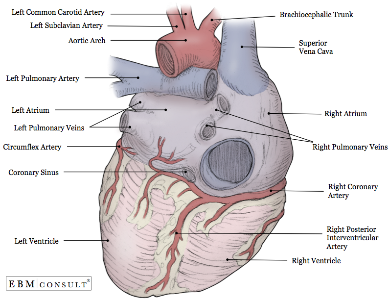
20864 Add to Lightbox. Aortc arch Ligamentum arteriosum Left pulmonary artery Left pulmonary ve ns Auricle of left atrium Circumflex artery Left coronary artery in atrioventricular sulcus Great cardiac vein Left ventricle Anterior interventricular artery in anterior interventricular sulcus Apex. One of these vessels the coronary sinus is returning to the right atrium the blood that has been to heart muscle SLIDES The next few slides focus on the coronary blood vessels.
The posterior view of the heart shows the prominent coronary surface vessels. Images on Similar Topics. Posterior View Choose a structure from the pull-down list.
Click on Label for the labeled model. What are the parts of the hearts anatomy. There is a printable worksheet available for download here so you can take the quiz with pen and paper.
Heart Anatomy Self Test. Back to Circulatory System. This is an online quiz called Label the Heart Posterior View.
8 branch to uppe. The heart sits within a fluid-filled cavity called the pericardial cavity. On average an adults heart weighs about 10 ounces.
1 1 summit of lungs. S branch to upper lobe of lung. Note that in most cases 23 of the heart is positioned to the left of midline and the.
The Heart Posterior View. 6 branch to lower lobe. Posterior View When you point to any structure on the photograph that region or structure will be highlighted in the smaller image to the left to help you locate it.
There is a printable worksheet available for download here so you can take the quiz with pen and paper. Denoyer- Geppert DG Medical Plastics Laboratory MPL Somso. Email this page.
Posterior structures of the heart Label the following structures in the posterior view of the heart. 62223 Add to Lightbox. Label the 4 chambers as well as the major vessels entering and leaving these chambers.
If you want to check your answers use the Reset Incorrect button. The parts of your heart are like the parts of a house. The left atrium left ventricle and coronary sinus in the coronary sulcus between these chambers can be easily seen from the posterior aspect.
Link this page. Click on the tags below to find other quizzes on the same subject. In this interactive you can label parts of the human heart.
The heart is shaped as a quadrangular pyramid and orientated as if the pyramid has fallen onto one of its sides so. If you click your left mouse button the name of that structure will appear to identify it. Up to 15 cash back Bronchi and lungs posterior view showing position of heart.
This illustration demonstrates a posterior view of the thoracic cavity highlighting the position of the heart in relationship to the ribs and diaphragm. The different blood vessels are labeled. Largest artery in the body.
Because the heart points to the left about 23 of the hearts mass is found on the left side of the body and the other 13 is on the right. 2 2 base of lungs. Your Skills Rank.
Posterior Aspect base of Heart With Light Micrograph of the Wall of the Superior Vena Cava And Light Micrograph of the Wall of the Inferior Vena Cava Variant Image ID. Start studying Heart Anatomy Posterior View. The top panel of this figure shows the anterior view of the heart while the bottom panel shows the posterior view of the heart.
Includes an exercise review worksheet quiz and model drawing of an anterior vi. Drag and drop the text labels onto the boxes next to the heart diagram. Then click on the matching structure in the large heart image.
Click on a photo for a larger view of the model. Carries deoxygenated blood from the right ventricle to the left lung. Anatomy of the Heart Pericardium.
Instant anatomy is a specialised web site for you to learn all about human anatomy of the body with diagrams podcasts and revision questions. If you want to redo an answer click on the box and the answer will go back to the top so you can move it to another box. Learn vocabulary terms and more with flashcards games and other study tools.
Your heart may weigh a little more or a little less depending on your body size and sex. This is an online quiz called Anatomy of the Human Heart - Posterior View. A Anterior view of the external heart C 2019 Pearson Education.
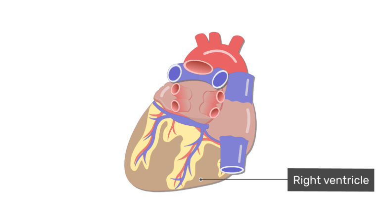
Function and anatomy of the heart made easy using labeled diagrams of cardiac structures and blood flow through the atria ventricles valves aorta pulmonary arteries veins superior inferior vena cava and chambers. Terms in this set 15 Aorta. Anterior view of heart focused on coronary vessels This view allows a student to observe the major coronary vessels of the heart.
Diseases of the Heart. Choose a Structure Here. With the 3D model a student can tilt and rotate the heart to get an optimal view of each coronary vessel.
The external anatomy of the heart has been set to Transparent but could easily be changed to Show.
Heart Posterior View Diagram Quizlet
The Heart Chambers And Their Functions
Anatomy Heart External

Posterior View Of The Heart Heart Anatomy Heart Diagram Anatomy

Heart Anatomy Anatomy And Physiology Ii

Posterior View Of The External Heart Diagram Quizlet

Posterior View Of The Heart Diagram Quizlet

Heart Anatomy Labelled Illustration Stock Image C043 4821 Science Photo Library
0 comments
Post a Comment